Bone Cross Section Anatomy : A Femur Cross Section Anatomy Of The Femur Cross Section Anatomy Medicine Com / Anatomies like brain, temporal bone/internal auditory meatus, nasopharynx, orbit, paranasal sinuses, cranial nerves, temporomandibular joint, neck, brachial plexus, spine, shoulder, arm, elbow, forearm, wrist, hand, finger, thumb, thorax/lung, coronary arteries, abdomen, pelvis, hip, thigh, knee, leg, ankle, foot, angiogram, etc.
Bone Cross Section Anatomy : A Femur Cross Section Anatomy Of The Femur Cross Section Anatomy Medicine Com / Anatomies like brain, temporal bone/internal auditory meatus, nasopharynx, orbit, paranasal sinuses, cranial nerves, temporomandibular joint, neck, brachial plexus, spine, shoulder, arm, elbow, forearm, wrist, hand, finger, thumb, thorax/lung, coronary arteries, abdomen, pelvis, hip, thigh, knee, leg, ankle, foot, angiogram, etc.. Related posts of cross section of a long bone foot bone anatomy x ray. A typical long bone shows the gross anatomical characteristics of bone. As we age, our bones lose their strength. It includes such bones as the hip and vertebrae. Two types of bone tissues in cross section of a long bone :
Antique illustration of human body anatomy bones, skull: For each level or axial section, there will be an actual ct brain image that is labeled and figures depicting where the scan is in the body. Foot bone anatomy x ray 12 photos of the foot bone anatomy x ray foot bone anatomy x ray, bone, foot bone anatomy x ray. A typical long bone shows the gross anatomical characteristics of bone. 12 photos of the cross section of human bone diagram.
Cross Sectional Anatomy Kenhub from thumbor.kenhub.com Antique illustration of human body anatomy bones, skull: There are trabeculae in spongy bone which gives its sponge like appearance. These canals are part of the osteon structure of the cortex. Download 706 bone cross medical section stock illustrations, vectors & clipart for free or amazingly low rates! 12 photos of the cross section of human bone diagram. Data and dicom images archived on the pacs (picture archiving and communicating system) were processed and exported as jpeg images. The central tubular region of the bone, called the diaphysis, flares outward near the end to form the metaphysis, which contains a largely cancellous, or spongy, interior. Cross section of a bone, this image shows the interior of the bone, which has a lot of spongy bone tissue.
This category is largely categorized by the content of the bone rather than the shape.
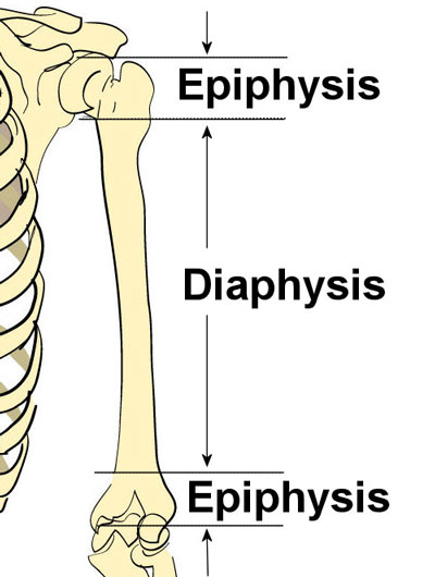
As we age, our bones lose their strength. They are obtained by taking imaginary slices perpendicular to the main axis of organs, vessels, nerves, bones, soft tissue, or even the entire human body. Anatomy of a flat bone. There are trabeculae in spongy bone which gives its sponge like appearance. This category is largely categorized by the content of the bone rather than the shape. Red marrow fills the spaces in the spongy bone. The wider section at each end of the bone is called the epiphysis (plural = epiphyses), which is filled with spongy bone. Bone on side of the foot Photomechanical print page item number: These canals are part of the osteon structure of the cortex. Internal structure of a human long bone, with a magnified cross section of the interior. It includes such bones as the hip and vertebrae. Histology sauropod vertebra picture of the week these pictures of this page are about:long bone cross section.
As the names suggest compact bone looks compact and the spongy bone looks like sponges. Photomechanical print page item number: They are obtained by taking imaginary slices perpendicular to the main axis of organs, vessels, nerves, bones, soft tissue, or even the entire human body. Synovial joint capsule bones chart. Antique illustration of human body anatomy bones, skull:
Bone Structure Anatomy Explained What Is Bone Marrow from www.teachpe.com The periosteum contains many strong collagen fibers that are used to firmly anchor tendons and muscles to the bone for movement. Looking at a bone in cross section, there are several distinct layered regions that make up a bone. Compact bone is the outer layer and the spongy bone forms the inner layer. Related posts of cross section of a long bone foot bone anatomy x ray. Bone matrix and cells bone matrix osseous tissue is a connective tissue and like all connective tissues contains relatively few cells and large amounts of extracellular matrix. Browse 4,287 bone cross section stock photos and images available, or search for human bone cross section to find more great stock photos and pictures. The outside of a bone is covered in a thin layer of dense irregular connective tissue called the periosteum. Data and dicom images archived on the pacs (picture archiving and communicating system) were processed and exported as jpeg images.
Bone is a specialised type of connective tissue.
Foot bone anatomy x ray 12 photos of the foot bone anatomy x ray foot bone anatomy x ray, bone, foot bone anatomy x ray. New users enjoy 60% off. Download 706 bone cross medical section stock illustrations, vectors & clipart for free or amazingly low rates! Cross section diagram of human bone, bone, cross section diagram of human bone. The compact bone is made up of osteon. As with other tools applied to petroleum development. It is to be hoped that a basic understanding of the anatomy of the temporal bone, and a systematic approach to the various pathologic entities that can affect it, can reduce the anxiety associated with, and improve. Compact bone is the outer layer and the spongy bone forms the inner layer. There are trabeculae in spongy bone which gives its sponge like appearance. Related posts of cross section of a long bone foot bone anatomy x ray. Bone matrix and cells bone matrix osseous tissue is a connective tissue and like all connective tissues contains relatively few cells and large amounts of extracellular matrix. For each level or axial section, there will be an actual ct brain image that is labeled and figures depicting where the scan is in the body. The outside of a bone is covered in a thin layer of dense irregular connective tissue called the periosteum.
Compact bone is the outer layer and the spongy bone forms the inner layer. Antique illustration of human body anatomy bones, skull: Anatomies like brain, temporal bone/internal auditory meatus, nasopharynx, orbit, paranasal sinuses, cranial nerves, temporomandibular joint, neck, brachial plexus, spine, shoulder, arm, elbow, forearm, wrist, hand, finger, thumb, thorax/lung, coronary arteries, abdomen, pelvis, hip, thigh, knee, leg, ankle, foot, angiogram, etc. It includes such bones as the hip and vertebrae. This category is largely categorized by the content of the bone rather than the shape.
Bone Structure Anatomy And Physiology I from s3-us-west-2.amazonaws.com As the names suggest compact bone looks compact and the spongy bone looks like sponges. For each level or axial section, there will be an actual ct brain image that is labeled and figures depicting where the scan is in the body. Learning cross sectional brain anatomy in this section, you will be presented with several images and figures illustrating the cross sectional brain anatomy that you will be learning. Browse 4,287 bone cross section stock photos and images available, or search for human bone cross section to find more great stock photos and pictures. Browse 4,275 bone cross section stock photos and images available, or search for human bone cross section to find more great stock photos and pictures. Cross section of a bone, this image shows the interior of the bone, which has a lot of spongy bone tissue. Antique illustration of human body anatomy bones, skull: Spongy bone and compact bone.
Compact bone is the outer layer and the spongy bone forms the inner layer.
Red marrow fills the spaces in the spongy bone. The central tubular region of the bone, called the diaphysis, flares outward near the end to form the metaphysis, which contains a largely cancellous, or spongy, interior. It is to be hoped that a basic understanding of the anatomy of the temporal bone, and a systematic approach to the various pathologic entities that can affect it, can reduce the anxiety associated with, and improve. It includes such bones as the hip and vertebrae. 12 photos of the cross section of human bone diagram. Foot bone anatomy x ray 12 photos of the foot bone anatomy x ray foot bone anatomy x ray, bone, foot bone anatomy x ray. Browse 4,275 bone cross section stock photos and images available, or search for human bone cross section to find more great stock photos and pictures. They are obtained by taking imaginary slices perpendicular to the main axis of organs, vessels, nerves, bones, soft tissue, or even the entire human body. Discover (and save!) your own pins on pinterest Antique illustration of human body anatomy bones, skull: New users enjoy 60% off. Photomechanical print page item number: For each level or axial section, there will be an actual ct brain image that is labeled and figures depicting where the scan is in the body.
Related posts of cross section of a long bone foot bone anatomy x ray bone cross section. This category is largely categorized by the content of the bone rather than the shape.


Komentar
Posting Komentar